


SEEFOR 17(1): 26015
Article ID: 26015
DOI: https://doi.org/10.15177/seefor.26-015
ORIGINAL SCIENTIFIC PAPER
Pathogenic Micromycete Neocatenulostroma germanicum in Pine Plantations of the Republic of Armenia
Anastasiia Shchukovskaya1, Elena Arbuzova1, Nataliia Kozyreva1, Karine Akopyan2, Tigran Ghrejyan2, Mark Kalashian2, Inessa Eloyan3, Iren Shahazizyan3, Gayane Karagyan2,*
Addresses:
(1) All-Russian Plant Quarantine Center, Pogranichnaya 32, RU-140150 Bykovo, Russia;
(2) National Academy of Sciences of the Republic of Armenia, Scientific Center of Zoology and Hydroecology, P. Sevak 7, AM-0014 Yerevan, Armenia;
(3) Yerevan State University, Faculty of Biology, Alek Manukyan 1/3, AM-0025 Yerevan, Armenia
Citation: Shchukovskaya A, Arbuzova E, Kozyreva N, Akopyan K, Ghrejyan T, Kalashian M, Eloyan I, Shahazizyan I, Karagyan G, 2026. Pathogenic Micromycete Neocatenulostroma germanicum in Pine Plantations of the Republic of Armenia. South-east Eur for 17(1): 26015. https://doi.org/10.15177/seefor.26-015 .
Received: 24 Sep 2025; Revised: 25 Feb 2026; Accepted: 12 Mar 2026; Published online: 29 Jun 2026
Cited by: Google Scholar
Abstract
From the needles of the Scots pine (Pinus sylvestris L.) collected in 2022 and 2023 in Northeastern and Central regions of the Republic of Armenia the causative agent of pine needles browning Neocatenulostroma germanicum (Crous & U. Braun) Quaedvlieg & Crous was identified. Both morphological and molecular approaches along with phylogenetic analysis were applied. This is the first report of N. germanicum identified in the territory of Armenia.
Keywords: ascomycetous fungus; pine needles browning; Pinus sylvestris; morphological examination; internally transcribed spacer (ITS) region; phylogenetic analysis
INTRODUCTION
Armenia has relatively limited forest resources. The total forest area in the country is approximately 11% of the total land area (Mkrtchyan and Grigoryan 2014, Bondarev 2020). Among these, pine plantations constitute the smallest share among the main forest-forming species, accounting for 5.3% of Armenia’s total forest area (Alaverdyan 2002).
Despite the relatively small area of pine forests in Armenia, they play an important economic and nature conservation role. Pine planting (Pinus spp.) in Armenia started in the mid-20th century primarily to replenish the forest fund in areas initially treeless or bare as a result of logging, in areas where pine was the most acceptable species for the existing soil and climatic conditions and where reforestation using other species was considered inappropriate. Currently, pine forests play an anti-erosion role on mountain slopes. They also have a certain protective value for agricultural lands, and contribute to the creation of favorable microclimatic conditions, particularly in areas of traditional recreation.
Drying of pine plantations, mainly artificially planted Pinus sylvestris, in Armenia was first observed in 2018 in the northern provinces. In the following years, this phenomenon spread widely to the central and southern regions of the country as well. To elucidate the underlying causes of this phenomenon, comprehensive surveys were carried out, including studies of insect pests (Karagyan et al. 2024) and nematodes (Arbuzova et al. 2025). Preliminary data on the fungal community were also obtained (Shchukovskaya et al. 2024).
In the frame of the reconnaissance surveys of fungal communities’ symptoms of lesion (browning and dying of the ends of needles), symptoms similar in appearance to those caused by Neocatenulostroma germanicum were found on needles of Pinus sylvestris L.
The genus Neocatenulostroma includes both plant-pathogenic species and those occurring on various substrates (Quaedvlieg et al. 2014). Within the genus, a distinct complex has formed, consisting of three morphologically similar and phylogenetically closely related species: N. abietis (Butin & Pehl) Quaedvlieg & Crous, N. germanicum (Crous & U. Braun) Quaedvlieg & Crous, and N. microsporum (Joanne E. Taylor & Crous) Quaedvlieg & Crous (Quaedvlieg et al. 2014, Markovskaja et al. 2016).
Recently, the identification of pathogenic micromycetes has increasingly relied on a combination of morphological and molecular diagnostic methods, which were also used in our study. This is the first record of this pathogenic species in the mycoflora of the country.
MATERIALS AND METHODS
Sample Collection
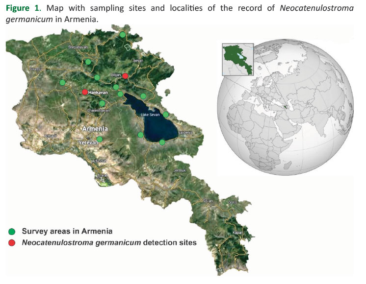
During 2022 and 2023, the surveys of drying pine plantations were carried out in some provinces of Armenia (Kotayk, Gegharkunik, Tavush, Lori and Aragatsotn) (Figure 1, Table 1). Samples were collected from 5 to 10 trees in each location, with the number of pine needles analyzed from each tree being approximately 15. Needles showing obvious signs of damage (such as discoloration, formation of spore-bearing structures, necrosis, and wilting) were collected. Wood fragments were collected for further mycological analyses as well.
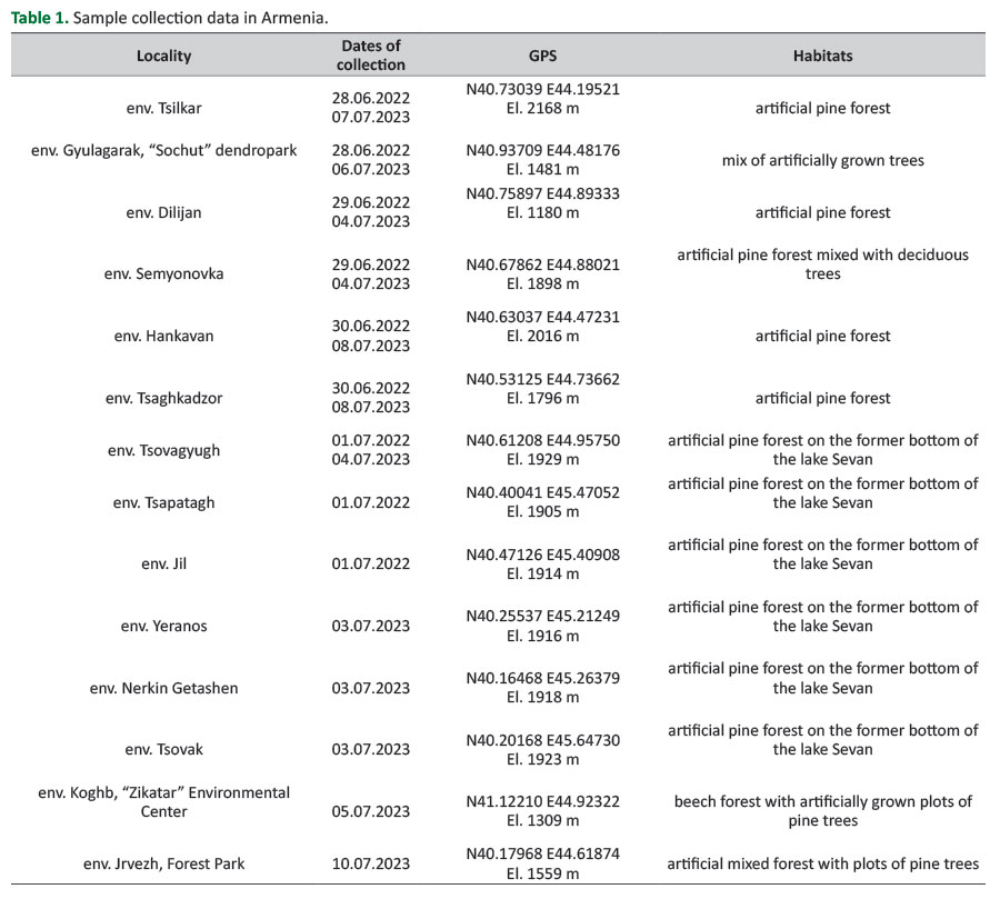 Table 1. Sample collection data in Armenia.
Fungi Isolation, Microscopy and Morphometry
Needle fragments showing signs of necrosis were sterilized in 70% ethanol for 10 seconds, then rinsed with sterile water 3–5 times, dried, placed in a moist chamber, and incubated for 1–2 weeks at 20–22°С (Stamets and Chilton 1983). The culture of the fungus was obtained by subculturing well-developed sporodochia that had formed on infected needles onto a nutrient medium potato dextrose agar (PDA). PDA medium was prepared from dry powder using the commercial “HiMedia Laboratories Pvt. Limited” (India) according to the manufacturer’s instruction. Fragments of mycelium for subculturing were taken from the periphery of actively growing colonies to avoid contamination by bacteria and other fungi. Isolation was also carried out on a growth medium (PDA), incubated for 3 to 6 weeks at the temperature of 20–22°С. For molecular analysis, a pure fungal culture was obtained through multiple subcultures on PDA.
The formed mycelium was examined under a light microscope Olympus BX53 Evident equipped with Olympus DP74 digital camera for revealing morphological structures. The sporodochia formed on the surface of the needles were photographed using a Zeiss SteReo Discovery V12 stereomicroscope equipped with a Canon EOS 5D MkIII camera. Initial species identification was carried out by morphological features using reference literature (Crous et al. 2007, Quaedvlieg et al. 2014, Markovskaja et al. 2016).
DNA Extraction, Polymerase Chain Reaction (PCR) Amplification and Sequencing
Genomic DNA was isolated from sporulating cultures using the commercial “PhytoSorb” kit (Syntol, Russia), based on purification with magnetic particles, in accordance with the manufacturer’s instructions. In the two studied isolates, the amplification of the nuclear genome region internal transcribed spacer (ITS) was performed using the following pair of universal primers (White et al. 1990):
ITS-5- 5’- GGAAGTAAAAGTCGTAACAAG G -3’,
ITS-4 - 5’- TCCTCCGCTTATTGATATGC -3’.
The PCR mixture (25 µl) contained 5 µl 5× PCR buffer MasOR TaqMix-2025 (“Dialat Ltd”, Russia), 0.5 µl of each primer, 2 µl of the respective genomic DNA extract and 17 µl sterile water. PCR conditions were as follows: initial denaturation for 3 min at 95°С, followed by 40 cycles of 30 s at 95°С, 30 s at 52°С and 30 s at 72°С, with final extension for 7 min at 72°С.
Visualization of PCR products was performed by gel electrophoresis in 1% agarose gel stained with ethidium bromide. PCR products intended for Sanger sequencing were purified using the GeneJET PCR Purification Kit (Thermo Fisher, USA) according to the manufacturer’s instructions.
The sequencing reaction was performed using BigDye Terminator v3.1 Cycle Sequencing Kit reagents (Applied Biosystems, USA) according to the manufacturer’s instructions, followed by separation of fragments on a 3500 genetic analyzer (Applied Biosystems, USA).
Two original sequences of N. germanicum specimens from Hankavan village (accession number PX219661) and Dilijan town (PX219657) obtained for the 572 bp fragment, and one sequence of Diplodia sapinea from Tsilkar village (PX219648) obtained for the 600 bp fragment of the ITS region were submitted to GenBank.
Samples and Sequence Alignment, Phylogenetic Tree Construction
Nucleotide sequences obtained in this study were edited and aligned using the Clustal W algorithm in the BioEdit software (Hall 1999). Comparative analysis of the obtained nucleotide sequences with the sequences deposited in GenBank was carried out using the BLAST program on the NCBI website (https://www.ncbi.nlm.nih.gov). Phylogenetic analysis was performed in MEGA 11.0 program (MEGA software development team, USA) using Maximum Likelihood (ML) method and Kimura 2-parameter model. The standard bootstrap (1000 replicates) was used to evaluate the statistical nodal support of the tree (Kimura 1980, Kumar et al. 2016).
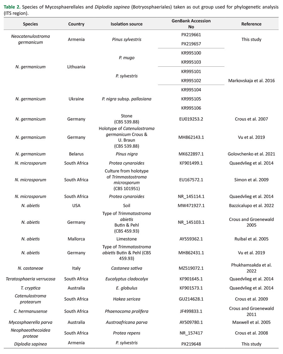
For phylogenetic analysis the ITS region sequences of 27 samples, both original and those obtained from GenBank were used, namely: 12 samples of Neocatenulostroma germanicum, 3 samples of N. microsporum, 4 samples of N. abietis, 1 sample of N. castaneae Phukhams., Bhunjun & K.D. Hyde, 6 species belonging to the other genera of order Mycosphaerellales and Diplodia sapinea (Fr.) Fuckel (Botryosphaeriales, as out group) (Table 2).
RESULTS AND DISCUSSION
Cultural and Morphological Characteristics of Neocatenul-ostroma Germanicum
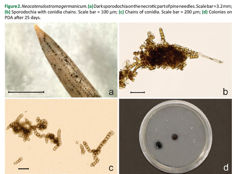
Laboratory studies on the surface of the necrotized part of the needles collected in the vicinity of Dilijan town (Tavush Province) and Hankavan village (Kotayk Province) revealed the formed conidial sporulation of the fungus – black stromatic beds, as well as sporodochia exiting through the stomata (Figures 2 a, b).
Conidia are thick-walled, dark brown, ellipsoid, with one septum, combined into short chains, 9.4–25.2 × 4.9–6.4 µm (Figure 2 c).
Colonies are slow-growing, on the 25th day at 20°С the average diameter was 11–12 mm, their shape is rounded, with smooth edges (Figure 2 d). At the initial stage of growth, the colony appears gray with an olive tinge; as it grows, it takes on a rich dark gray (steel-like) color with a faint olive-green tint. The colony reverse is uniformly black in color.
According to morphological characteristics, the isolated fungus was identified as Neocatenulostroma germanicum (Crous et al. 2007, Markovskaja et al. 2016).
Comparison of the Sequences and Phylogenetic Analysis
According to Maximum Likelihood (ML) phylogenetic analysis of the original sequences obtained in this study and deposited in GenBank, some sequences showed that they belong to the Neocatenulostroma abietis/germanicum/microsporum species complex (Figure 3). As expected, these species form a single clade with high support (99%) of the bootstrap test, whereas the newly described species N. castaneae from Italy (MZ519072.1) is not included in this clade. It should be noted that the target isolates from Armenia are 99.6–99.8% identical to N. germanicum isolates from Germany (EU019253.2), Latvia (KR995101), and Ukraine (KR995104, KR995105, KR995106).
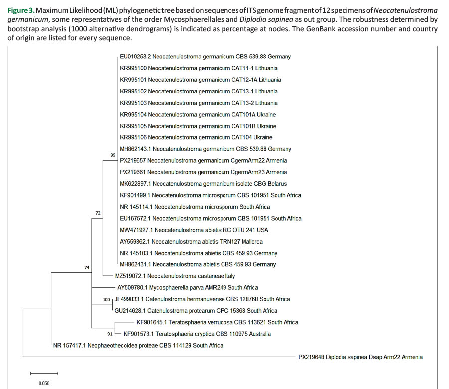
Among morphologically similar species of the group, which include Neocatenulostroma abietis, N. germanicum and N. microsporum, one of the most potentially dangerous and poorly studied pathogenic micromycetes is N. germanicum, the causal agent of needle browning. The species is distributed in several European countries, including Germany, Poland, Lithuania, Montenegro, Belarus, and some regions of Russia (Markovskaja et al. 2016, Lasarević and Menkis 2020, Golovchenko et al. 2021, Bulgakov 2022). N. germanicum can behave not only as a saprotroph developing on various substrates, but also as a serious pathogen, massively affecting the needles of young pines in spring and summer, sometimes together with fungi of the genus Dothiostroma sp. (Markovskaja et al. 2016). Therefore, the application of both microscopic and morphometric methods, as well as molecular analysis (using a part of ITS region) confirmed the presence of N. germanicum in two localities of the artificial pine plantations in the vicinity of the Dilijan town and the Hankavan village, in the Republic of Armenia. In both localities, artificially grown pine forests planted on fairly steep, south-facing slopes are present. Despite different altitudes, the habitats are characterized by similar ecological conditions.
CONCLUSION
The results of our research allow us to draw a definite conclusion that in the north-eastern and central regions of the Republic of Armenia (Tavush and Kotayk provinces) on the needles of Scots pine (Pinus sylvestris L.) a pathogenic micromycete Neocatenulostroma germanicum has been revealed, whose species identity is confirmed by the results of studies based on morphological, microscopic and molecular diagnostic methods. This is the first report of the discovery of the species on the territory of the Republic of Armenia.
It is important to note that further molecular genetic studies are planned, during which more variable and informative regions of the nuclear genome will be tested to enable more precise species differentiation within the genus Neocatenulostroma.
Author Contributions
KG, GT – conceived and designed the research, KG, AK, GT, SA, AE, EI, SI – conducted field research and collected material, SA, KN, – performed laboratory studies, SA, KN, KG – processed the data, KG - secured the field research funding; KG, KM – assisted with the preparation of the drafted manuscript, SA – wrote the manuscript.
Funding
The work of coauthors from Armenia was supported by the Higher Education and Science Committee of MESCS RA, in the frames of the research project № 20TTWS-1F017 and partially project № 24WS-1F030.
Acknowledgments
We would like to express our gratitude to the staff of the “Sevan” and “Dilijan” National Parks and the “Zikatar” State Sanctuary, as well as some forestry enterprises of Armenia for their assistance in carrying out our field surveys.
Conflicts of Interest
The authors declare no conflict of interest.
REFERENCES
Alaverdyan AZ, 2002. Forest formation of Pinus hamata (Steven) Sosn. in Ijevan Region. Takhtajania 14: 103-104. (in Russian).
Arbuzova EN, Karagyan GH, Kozyreva NI, Shchukovskaya AG, Ghrejyan TL, Kalashian MYu, Akopyan KV, 2025. First finding of Bursaphelenchus xylophilus in pine plantations of the Republic of Armenia. J Nematol 57 (1): 1-10. https://doi.org/10.2478/jofnem-2025-0004.
Bazzicalupo AL, Erlandson S, Branine M, Ratz M, Ruffing L, Nguyen NH, Branco S, 2022. Fungal community shift along steep environmental gradients from geothermal soils in Yellowstone National Park. Microb Ecol 84(1): 33-43. https://doi.org/10.1007/s00248-021-01848-y.
Bondarev A, 2020. National Strategy for the inventory of Forests in the Republic Armenia. Ministry of Enviroment of the Repiblic of Armenia, Yerevan.
Bulgakov TS, 2022. About poor-known dangerous diseases of pine needles in Southern Russia. Actual Issues of Forest Complex 61: 74-79.
Crous PW, Braun U, Groenewald JZ, 2007. Mycosphaerella is polyphyletic. Stud Mycol 58: 1-32. https://doi.org/10.3114/sim.2007.58.01.
Crous PW, Groenewald JZ, 2005. Hosts, species and genotypes: opinions versus data. Australasian Plant Pathol 34: 463–470. https://doi.org/10.1071/AP05082.
Crous PW, Groenewald JZ, 2011. Why everlastings don't last. Persoonia 26: 70-84. https://doi.org/10.3767/003158511X574532.
Crous PW, Schoch CL, Hyde KD, Wood AR, Gueidan C, de Hoog GS, Groenewald JZ, 2009. Phylogenetic lineages in the Capnodiales. Stud Mycol 64: 17-47. https://doi.org/10.3114/sim.2009.64.02.
Crous PW, Summerell BA, Mostert L, Groenewald JZ, 2008. Host specificity and speciation of Mycosphaerella and Teratosphaeria species associated with leaf spots of Proteaceae. Persoonia 20: 59–86. https://doi.org/10.3767/003158508X323949.
Golovchenko LA, Dishuk NG, Panteleev SV, Baranov OYu, 2021. New data of the spread of the invasive species Dothiostroma septosporum (Dorogin) M. Morelet in Belarus. Proceedings of the National Academy of Sciences of Belarus Biological series 66(2): 147-158. https://doi.org/10.29235/1029-8940-2021-66-2-147-158.
Hall TA, 1999. BioEdit: a user-friendly biological sequence alignment editor and analysis program for Windows 95/98/NT. Nucl Acid S 41: 95–98.
Karagyan GH, Ghrejyan TL, Arbuzova EN, Akopyan KV, Shchukovskaya AG, Shahazizyan IV, 2024. Quarantine, invasive and expansive invertebrates and phytopathogenic mycoflora of pine plantings of Armenia. Plant Health and Quar 1 S (18): 95-96.
Kimura MA, 1980. Simple method for estimating evolutionary rate of base substitutions through comparative studies of nucleotide sequences. J Mol Evol 16: 111-120. https://doi.org/10.1007/bf01731581.
Kumar S, Stecher G, Tamura K, 2016. MEGA7: Molecular Evolutionary Genetic Analysis version 7.0 for bigger datasets. Mol Biol Evol 33: 1870-1874. https://doi.org/10.1093/molbev/msw054.
Lasarević J, Menkis A, 2020. Fungal Diversity in the Phyllosphere of Pinus heldreichii H. Christ – An Endemic and High-Altitude Pine of the Mediterranean Region. Diversity 12(5): 172. https://doi.org/10.3390/d12050172.
Lasarević J, Menkis A, 2022. Cytospora friesii and Sydowia polyspora are associated with the sudden dieback of Abies concolor in Southern Europe. Plant Prot Sci 58 (3): 258–263. https://doi.org/10.17221/120/2021-PPS.
Markovskaja S, Kačergius A, Davydenko K, Fraser S, 2016. First record of Neocatenulostroma germanicum on pines in Lithuania and Ukraine and its co-occurrence with Dothistroma spp. and other pathogens. For Pathol 46: 522-533. https://doi.org/10.1111/efp.12308.
Maxwell A, Jackson SL, Dell B, Hardy GE, 2005. PCR-identification of Mycosphaerella species associated with leaf diseases of Eucalyptus. Mycol Res 109(9): 992-1004. https://doi.org/10.1017/S0953756205003539.
Mkrtchyan A, Grigoryan E, 2014. Forest Dependency in Rural Armenia. FLEG II (ENPI East) Program/World Bank, Yerevan, Armenia.
Phukhamsakda C, Nilsson RH, Bhunjun CS, de Farias ARG, Sun Y-R, Wijesinghe SN, Raza M, Bao D-F, Lu L, Tibpromma S, Dong W, Tennakoon DS, Tian X-G, Xiong Y-R, Karunarathna SC, Cai L, Luo Z-L, Wang Y, Manawasinghe IS, Camporesi E, Kirk PM, Promputtha I, Kuo C-H, Su H-Y, Doilom M, Li Y, Fu Y-P, Kevin DH, 2022. The numbers of fungi: contributions from traditional taxonomic studies and challenges of metabarcoding. Fungal Divers 114: 327-386. https://doi.org/10.1007/s13225-022-00502-3.
Quaedvlieg W, Binder M, Groenewald JZ, Summerell BA, Carnegie AJ, Burgess TI, Crous PW, 2014. Introducing the Consolidated Species Concept to resolve species in the Teratosphaeriaceae. Persoonia 33: 1-40. https://doi.org/10.3767/003158514X681981.
Ruibal C, Platas G, Bills G, 2005. Isolation and characterization of melanized fungi from limestone formations in Mallorca. Mycol Prog 4: 23–38. https://doi.org/10.1007/s11557-006-0107-7.
Shchukovskaya A, Arbuzova E, Karagyan G, Akopyan K, Ghrejyan T, Shahazizyan I, 2024. Study of invasive and quarantine phytopathogenic fungi of pine plantations of the Republic of Armenia. Plant Health and Quar 2 SA 18B: 62-63 (In Russian).
Simon UK, Groenewald JZ, Crous PW, 2009. Cymadothea trifolii, an obligate biotrophic leaf parasite of Trifolium, belongs to Mycosphaerellaceae as shown by nuclear ribosomal DNA analyses. Persoonia 22: 49-55. https://doi.org/10.3767/003158509X425350.
Stamets P, Chilton JS, 1983. The mushroom cultivator: a practical guide to growing mushrooms at home. Agarikon Press, Olympia, WA, USA, 415 p.
Vu D, Groenewald M, de Vries M, Gehrmann T, Stielow B, Eberhardt U, Al-Hatmi A, Groenewald JZ, Cardinali G, Houbraken J, Boekhout T, Crous PW, Robert V, Verkley GJM, 2019. Large-scale generation and analysis of filamentous fungal DNA barcodes boosts coverage for kingdom fungi and reveals thresholds for fungal species and higher taxon delimitation. Stud Mycol 92: 135-154. https://doi.org/10.1016/j.simyco.2018.05.001.
White TJ, Bruns TD, Lee SB, Taylor JW, 1990. Amplification and direct sequencing of fungal ribosomal RNA genes for phylogenetics. In: Innis MA, Gelfand DH, Sninsky JJ, White TJ (eds) PCR protocols: a guide to methods and applications. Academic Press, New York, USA, 315-322. http://dx.doi.org/10.1016/B978-0-12-372180-8.50042-1.
© 2026 by the author(s). License: Croatian Forest Research Institute. This article is an open access article distributed under the terms and conditions of the Creative Commons Attribution 4.0 International License (CC BY 4.0).